هدف: ارائه روش های اتوماتیک و نیمه اتوماتیک برای کمک به افزایش دقت در تشخیص بیماری های چشم در انواع تصاویر چشم
- مدلسازی
- پیش پردازش
- بخش بندی (segmentation)
- طبقه بندی (classification)
- تشخیص ناهنجاری
- ارزیابی تقارن
- رجیستریشن
نقشه راه

- مقالات چاپ شده در مرکز مرتبط با موضوع مدل کردن آماری ، هندسی و انرژی دیتا در انواع تصاویر چشم
. Jorjandi, Z. Amini, G. Plonka, H. Rabbani, “Statistical modeling of retinal optical coherence tomography using the Weibull mixture model,” Biomedical Optic Express, 2021
. Samieinasab, Z. Amini and H. Rabbani, "Multivariate Statistical Modeling of Retinal Optical Coherence Tomography,", IEEE Transactions on Medical Imaging, 2020
. Amini and H. Rabbani, "Statistical Modeling of Retinal Optical Coherence Tomography," IEEE Transactions on Medical Imaging, 2016
. Tajmirriahi, Z. Amini, A. Hamidi, A. Zam and H. Rabbani, "Modeling of Retinal Optical Coherence Tomography Based on Stochastic Differential Equations: Application to Denoising," IEEE Transactions on Medical Imaging, 2021
. Monemian, H. Rabbani, “Mathematical analysis of texture indicators for the segmentation of optical coherence tomography images,” Optik, 2020
. Amini, H. Rabbani and I. Selesnick, "Sparse Domain Gaussianization for Multi-Variate Statistical Modeling of Retinal OCT Images," IEEE Transactions on Image Processing, 2020
- مقالات چاپ شده در مرکز مرتبط با موضوع آماده سازی تصویر از طریق کاهش نویز، بهبود کنتراست و ... برای پردازش های اصلی
Ezhei, G. Plonka, and H. Rabbani, "Retinal optical coherence tomography image analysis by a restricted Boltzmann machine," Biomedical Optic Express, 2022.
Esmaeili, A. M. Dehnavi, F. Hajizadeh, H. Rabbani, "Three-dimensional curvelet-based dictionary learning for speckle noise removal of optical coherence tomography," Biomedical Optic Express, 2020.
Kafieh, H. Rabbani and G. Unal, "Bandlets on Oriented Graphs: Application to Medical Image Enhancement," IEEE Access, 2019.
Tajmirriahi, R. Kafieh, Z. Amini and H. Rabbani, "A Lightweight Mimic Convolutional Auto-Encoder for Denoising Retinal Optical Coherence Tomography Images," IEEE Transactions on Instrumentation and Measurement, 2021.
Amini, H. Rabbani, “Optical coherence tomography image de-noising using Gaussianization transform,” Journal of Biomedical Optics, 2017.
- مقالات چاپ شده در مرکز مرتبط با موضوع تعیین مرز لایه های شبکیه یا مرز ناهنجاریهای شبکیه
M. Montazerin, Z. Sajjadifar, E. Khalili Pour, et al. ”Livelayer: a semi-automatic software program for segmentation of layers and diabetic macular edema in optical coherence tomography images,” Scientific Reports, 2021.
A. Rashno, B. Nazari, D. D. Koozekanani, P. M. Drayna, S. Sadri, H. Rabbani, K. K. Parhi, “Fully-automated segmentation of fluid regions in exudative age-related macular degeneration subjects: Kernel graph cut in neutrosophic domain,” PLOS ONE, 2017.
R. Kafieh, S. Rakhshani, J. Hogg, R. A. Lawson, N. Pavese, W. Innes, et al. “A robust, flexible retinal segmentation algorithm designed to handle neuro-degenerative disease pathology (NDD-SEG),” Investigative Ophthalmology & Visual Science, 2022.
- مقالات چاپ شده در مرکز مرتبط با موضوع دسته بندی تصاویر بر اساس معیارهای مختلف
L. Huang, X. He, L. Fang, H. Rabbani and X. Chen, "Automatic Classification of Retinal Optical Coherence Tomography Images With Layer Guided Convolutional Neural Network," IEEE Signal Processing Letters, 2019.
L. Fang, C. Wang, S. Li, H. Rabbani, X. Chen and Z. Liu, "Attention to Lesion: Lesion-Aware Convolutional Neural Network for Retinal Optical Coherence Tomography Image Classification," IEEE Transactions on Medical Imaging, 2019.
R. Rasti, H. Rabbani, A. Mehridehnavi and F. Hajizadeh, "Macular OCT Classification Using a Multi-Scale Convolutional Neural Network Ensemble," IEEE Transactions on Medical Imaging, 2018.
L. Fang, C. Wang, S. Li, J. Yan, X. Chen, H. Rabbani, “Automatic classification of retinal three-dimensional optical coherence tomography images using principal component analysis network with composite kernels,” Journal of Biomedical Optics, 2017.
E. Mousavi, R. Kafieh, H. Rabbani, “Classification of dry age-related macular degeneration and diabetic macular edema from optical coherence tomography images using dictionary learning,” IET image processing, 2020.
- مقالات چاپ شده در مرکز مرتبط با موضوع تشخیص ناهنجاری ها در تصاویر OCT و فندوس
L. Fang, C. Wang, S. Li, H. Rabbani, X. Chen and Z. Liu, "Attention to Lesion: Lesion-Aware Convolutional Neural Network for Retinal Optical Coherence Tomography Image Classification," IEEE Transactions on Medical Imaging, 2019.
M. Monemian, H. Rabbani, “Red-lesion extraction in retinal fundus images by directional intensity changes’ analysis,” Scientific Reports, 2021.
M. Monemian, H. Rabbani, “Directional analysis of intensity changes for determining the existence of cyst in optical coherence tomography images,” Scientific Reports, 2022.
Z. Baharlouei, H. Rabbani and G. Plonka, "Detection of Retinal Abnormalities in OCT Images Using Wavelet Scattering Network," 2022 44th Annual
International Conference of the IEEE Engineering in Medicine & Biology Society (EMBC), Glasgow, Scotland, United Kingdom, 2022, pp. 3862-3865,
doi: 10.1109/EMBC48229.2022.9871989.
- مقالات چاپ شده در مرکز مرتبط با موضوع بررسی تقارن بین تصاویر مختلف چشم چپ و راست
M. Mokhtari, H. Rabbani, A. M. Dehnavi, R. Kafieh, M. R. Akhlaghi, M. Pourazizi, L. Fang, “Local comparison of cup to disc ratio in right and left eyes based on fusion of color fundus images and OCT B-scans,” Information Fusion, 2019.
T. Mahmoudi, R. Kafieh, H. Rabbani, “Evaluation of asymmetry in right and left eyes of normal individuals using extracted features from optical coherence tomography and fundus images,” Journal of Medical Signals and Sensors, 2020.
- مقالات چاپ شده در مرکز مرتبط با موضوع رجیستریشن
E. Golkar, H. Rabbani, and A. Dehghani, "Hybrid registration of retinal fluorescein angiography and optical coherence tomography images of patients with diabetic retinopathy," Biomedical Optic Express, 2021.
R. Almasi, A. Vafaei, Z. Ghasemi, M. R. Ommani, A. R. Dehghani, H. Rabbani, “Registration of fluorescein angiography and optical coherence tomography images of curved retina via scanning laser ophthalmoscopy photographs,” Biomedical Optic Express, 2020.
- تمرکز بر تصاویر OCT A (برنامه های آتی مرکز)
Hojati, R. Kafieh, P. Fardafshari, M. Aghsaei Fard, H. Fouladi, “A MATLAB package for automatic extraction of flow index in OCT-A images by intelligent vessel manipulation,” SoftwareX, 2020.
- پروژه های جاری
•تشخیص بیماری DR و تعیین شدت آن
•بررسی تقارن چشم چپ و راست
•جمع آوری دیتاست های لازم از سراسر کشور و خارج از کشور
- دیتاست های جمع آوری شده (دانلود)
OCT data and color fundus images of left and right eyes of 50 healthy persons
Fundus fluorescein angiogram photographs of diabetic patients
Fundus fluorescein angiogram photographs and color fundus images of diabetic retinopathy
Color fundus images of healthy persons and patients with diabetic retinopathy
Database of 22 retinal images for the purpose of vessel-based registration of fundus and OCT projection images of retina
Dataset for OCT classification (50 Normal, 48 AMD & 50 DME)
ONH-based OCT of 7 healthy and 7 Glaucoma data captured by Heidelberg Spectralis
نرم افزارهای تولید شده
- نرم افزار تشخیص لکه های قرمز از تصاویر فندوس و رجیستریشن آنها

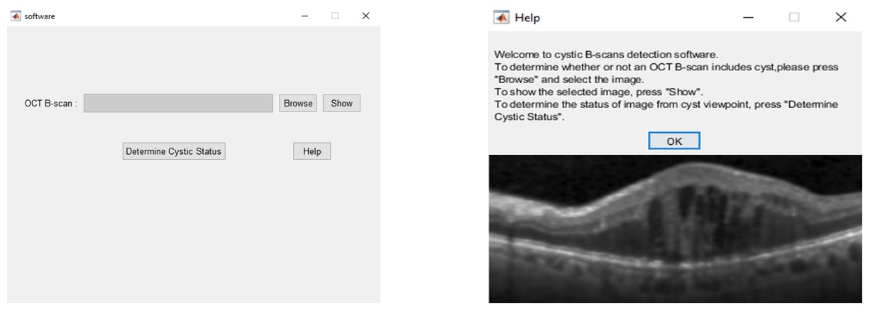
- نرم افزار تشخیص OCT B-scan های حاوی کیست

جلسات هسته چشم:
جلسات هسته چشم چهارشنبه هرهفته ساعت 9 صبح به صورت حضوری و مجازی برگزار می گردد.
دانلود فایل جلسه اول: 30 شهریور
طرح های تحقیقاتی در حال انجام در این حوزه
| عنوان طرح | نام مجری طرح | وضعیت فعلی طرح |
| استفاده از نشانگرهای پیشرفته بافت برای تشخیص میکروآنوریسم در تصاویر FA شبکیه | دکتر مریم منعمیان | در حال اجرا |
| تشخیص بیماری های شبکیه چشم از روی تصاویر مقطع نگاری همدوسی با استفاده از تبدیل پراکندگی موجک | دکتر زهرا بهارلویی | در حال اجرا |
| دسته بندی تصاویر OCT-A شبکیه چشم به دو حالت سالم و دیابتی با کمک ویژگی های آماری و تبدیل کرولت | دکتر مریم منعمیان | در حال اجرا |
| تشخیص hyper reflective foci از تصاویر OCT شبکیه با کمک تبدیل کرولت | دکتر مریم منعمیان | در حال اجرا |
جهت کسب اطلاعات بیشتر با شماره 031-37925252 تماس حاصل فرمایید.

