OCT data & Color Fundus Images of Left & Right Eyes of 50 healthy persons This dataset contains OCT data (in mat format) and color fundus data (in jpg format) of left & right eyes of 50 healthy persons. |
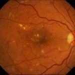
Fundus Fluorescein Angiogram Photographs of Diabetic Patients We have collected retinal image of 70 patients of different diabetic retinopathy stages including 30 normal data and 40 abnormal data in different stages. |
Database of corneal OCT taken from Heidelberg OCT imaging system (3D .mat data of 15 subjects) A set of 2D .mat corneal OCT images of 15 subjects. For example subject#1 includes 41 240×748 B-scans taken from Heidelberg OCT imaging device. |
Dataset for Fluorescein Angiography (Video & Late Image) in DME eyes The datasets (24 768*768*x FA videos and late FA images in DME eyes) and manual and automated markings used in the following paper can be downloaded from HERE. |
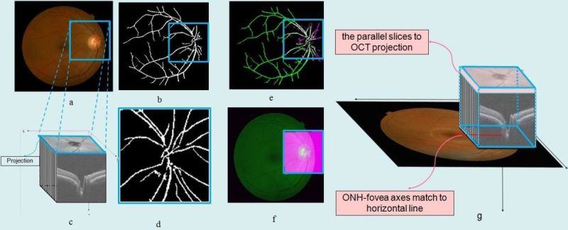
Database of 22 retinal images for the purpose of vessel-based registration of Fundus and OCT projection images of retina A set of eye images consisting of 22 pairs of images (17 macular and 5 prepapillary), from random patients, each pair acquired from eyes with a variety of retinal diseases. |
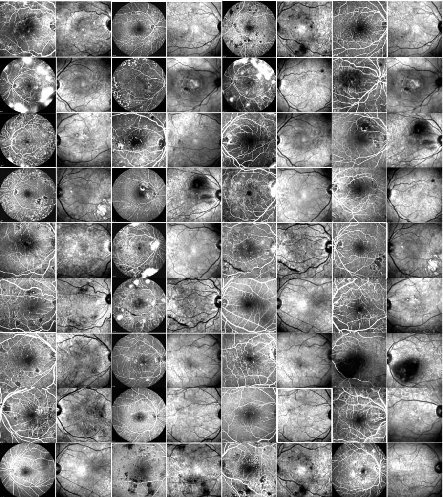
Fundus Fluorescein Angiogram Photographs & Colour Fundus Images of Diabetic Patients Publicly available database of both fundus fluorescein fngiogram photographs and corresponding color fundus images of 30 healthy persons and 30 patients with diabetic retinopathy. |
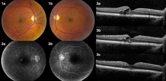
ONH-based OCT of 7 healthy and 7 glaucoma data captured by Heidelberg Spectralis 7 healthy and 7 glaucoma data captured by Heidelberg Spectralis used to demonstrate the efficacy of a new imaging biomarker namely Volumetric Cup-to-Disc Ratio (VCDR) for diagnosis of ocular diseases such as Glaucoma. |
Colour Fundus Images of Healthy Persons & Patients with Diabetic Retinopathy This folder includes 25 colour fundus images of healthy persons and 35 colour fundus images of patients with diabetic retinopathy used for automatic curvelet-based detection of Foveal Avascular Zone (FAZ). |
FA and SLO images of 21 subjects with diabetic retinopathy captured via Heidelberg Spectralis HRA2/OCT device This dataset contains 36 pairs of FA and SLO images of 21 subjects with diabetic retinopathy in jpg format are captured via Heidelberg Spectralis HRA2/OCT device and used for automatic registration. FA images were captured with two different fields of views (30 and 55 degrees). |
Dataset for OCT Classification (50 Normal, 48 AMD & 50 DME) This dataset is acquired at Noor Eye Hospital in Tehran and is consisting of 50 normal, 48 dry AMD, and 50 DME OCTs. |
The data of this dataset was acquired from a Custom-made swept-source OCT (SS-OCT) imaging system designed and built in Dep. of Biomedical Engineering, University of Basel.
Read More |
Topcon 3D-OCT Diabetic Data for Denoising This dataset contains six 3D OCT data using Topcon 3D OCT-1000 imaging system in Ophthalmology Dept., Feiz Hospital, Isfahan, Iran . The datasets are in mat format and are named “1” to “6”. Subjects in the dataset were diagnosed to have retinal Pigment Epithelial Detachment (PED).
|
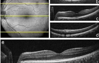
MA and normal strips of 20 subjects with diabetic retinopathy captured via Heidelberg device
|
Thirteen 3D macular SD-OCT images obtained from eyes without pathologies using Topcon 3D OCT-1000 imaging system This dataset contains thirteen 3D macular SD-OCT images obtained from eyes without pathologies using Topcon 3D OCT-1000 imaging system in Ophthalmology Dept., Feiz Hospital, Isfahan, Iran. |
Software for Red-lesion extraction in retinal fundus images by directional intensity changes’ analysis
This software has been prepared for staining in Fundus images as well as registering Fundus images. |
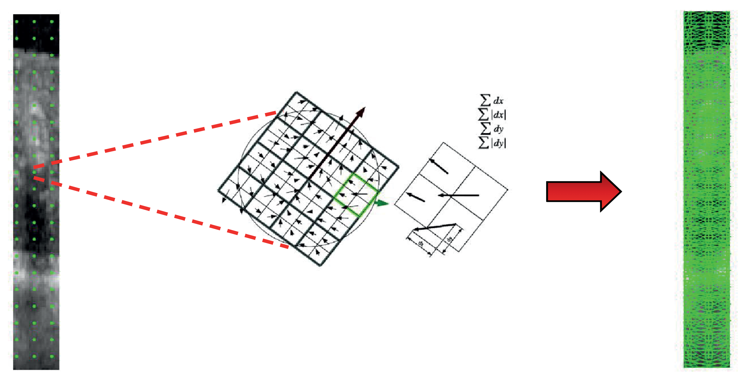
Four-class dataset of OCT image strips in PNG format With the help of related FAs, the OCT strips are created from OCT B-scans in four labels including MA, normal, abnormal, and vessel. Here, two datasets are prepared |
| Angio-Fundus |
Dataset of Fully-labeled Diabetic Macular Edema OCT B-scans (associated with fluid and layer annotations) We enrolled twenty eyes from 19 patients with the diagnosis of diabetic macular edema (DME). All patients had clinical and OCT-based diagnosis of DME. OCT examinations were performed using Spectralis Spectral Domain-OCT for .the normal and DME patients |
| Color Fundus Images with Exudates | Color Fundus Images |
Comprehensive Topcon 3D-OCT Dataset: Inclusive of Normal and AbnormalOCT Volumes |