|
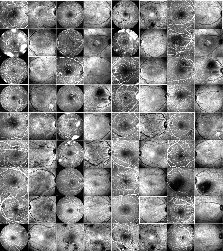
Fundus Fluorescein Angiogram Photographs of Diabetic Patients We have collected retinal image of 70 patients of different diabetic retinopathy stages including 30 normal data and 40 abnormal data in different stages. |
Bone Marrow Microscopic Data (plasma cell lineage images) This folder contains bone marrow microscopic images. These images are categorized into two groups: Normal Plasma Cells and Myeloma Cells. |
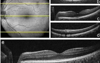
OCT data & Color Fundus Images of Left & Right Eyes of 50 healthy persons This dataset contains OCT data (in mat format) and color fundus data (in jpg format) of left & right eyes of 50 healthy persons. |
Dataset for Fluorescein Angiography (Video & Late Image) in DME eyes The datasets (24 768*768*x FA videos and late FA images in DME eyes) and manual and automated markings used in the following paper can be downloaded from HERE. |
|
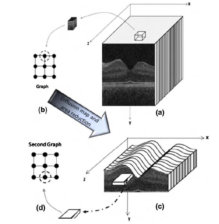
Database of corneal OCT taken from Heidelberg OCT imaging system (3D .mat data of 15 subjects) A set of 2D .mat corneal OCT images of 15 subjects. For example subject#1 includes 41 240×748 B-scans taken from Heidelberg OCT imaging device. |
Fundus Fluorescein Angiogram Photographs & Colour Fundus Images of Diabetic Patients Publicly available database of both fundus fluorescein fngiogram photographs and corresponding color fundus images of 30 healthy persons and 30 patients with diabetic retinopathy. |
CT & MR Volumes Used for Watermarking of DICOM Images This dataset contains 260 CT and 202 MR images in DICOM format. |
Dataset of Leishmania Parasite in Microscopic Images 45 24-bit 3264*2448 microscopic images taken from bone marrow samples including leshman bodies. |
|
Kidney microscopic images (Glomeruli) A dataset for Glomeruli detection was collected with the contribution of MISP Research Center and Department of Pathology at IUMS |
Red blood cells A self-provided dataset contains 148 microscopic images of blood smears. |
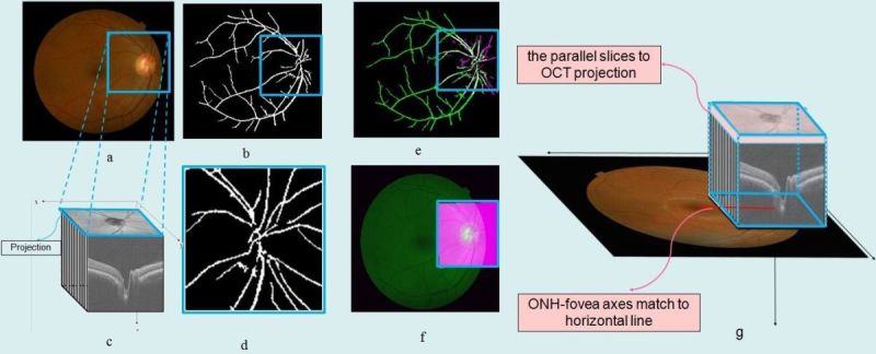
Database of 22 retinal images for the purpose of vessel-based registration of Fundus and OCT projection images of retina A set of eye images consisting of 22 pairs of images (17 macular and 5 prepapillary), from random patients, each pair acquired from eyes with a variety of retinal diseases. |
Colour Fundus Images of Healthy Persons & Patients with Diabetic Retinopathy This folder includes 25 colour fundus images of healthy persons and 35 colour fundus images of patients with diabetic retinopathy used for automatic curvelet-based detection of Foveal Avascular Zone (FAZ). |
|
Cardiac MRI short axis (SA) Cardiac MRI (CMRI), the images of the left ventricular region were selected for all frames and their contrast was increased by windowing.all slices are segmented interactively exactly based on the algorithm introduced by Heiberg. |
ONH-based OCT of 7 healthy and 7 glaucoma data captured by Heidelberg Spectralis 7 healthy and 7 glaucoma data captured by Heidelberg Spectralis used to demonstrate the efficacy of a new imaging biomarker namely Volumetric Cup-to-Disc Ratio (VCDR) for diagnosis of ocular diseases such as Glaucoma. |
Dataset for OCT Classification (50 Normal, 48 AMD & 50 DME) This dataset is acquired at Noor Eye Hospital in Tehran and is consisting of 50 normal, 48 dry AMD, and 50 DME OCTs. |
EEG Signals From Normal and MCI ( Mild Cognitive Impairment ) Cases This dataset is a collection of scalp EEG from 27 subjects ( 16 normal and 11 MCI ) aged 60 to 77 with elementary or higher education and history of coronary angiography during recent year. |
|
FA and SLO images of 21 subjects with diabetic retinopathy captured via Heidelberg Spectralis HRA2/OCT device This dataset contains 36 pairs of FA and SLO images of 21 subjects with diabetic retinopathy in jpg format are captured via Heidelberg Spectralis HRA2/OCT device and used for automatic registration. FA images were captured with two different fields of views (30 and 55 degrees). |
Voice Samples of Patients with Parkinson’s disease (spontaneous swallows in Parkinson’s disease) Data were collected from 34 subjects (19 males) who had Parkinson’s disease (PD) (age = 59.85 ± 11.46 years). They were referred for video fluoroscopy swallow study (VFSS) assessment as part of their routine medical care. |
Voice Samples of Patients with Internal Nasal Valve Collapse Before and After Functional Rhinoplasty This dataset contains voice samples of Patients with Internal Nasal Valve Collapse Before and After Functional Rhinoplasty. These voice samples are categorized into two groups: before and after functional rhinoplasty in patients with internal nasal valve collapse. |
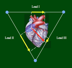
Vectorcardiography ( VCG ) The sampling rate was 500 Hz, and the samples were typically gathered for 16- second duration. The recorder device was Cardiax recorder. The 12-leads ECG and VCG signals were used in this study. each number in text file corresponds the leads except avr , all and avf. |
|
Topcon 3D-OCT Diabetic Data for Denoising This dataset contains six 3D OCT data using Topcon 3D OCT-1000 imaging system in Ophthalmology Dept., Feiz Hospital, Isfahan, Iran . The datasets are in mat format and are named “1” to “6”. Subjects in the dataset were diagnosed to have retinal Pigment Epithelial Detachment (PED).
|
OCT Basel Data The data of this dataset was acquired from a Custom-made swept-source OCT (SS-OCT) imaging system designed and built in Dep. of Biomedical Engineering, University of Basel. The central wavelength, spectral bandwidth and A-scan rate of the custom-made SS-OCT are 1064 nm, 100 nm, and 100 kHz, respectively.
|
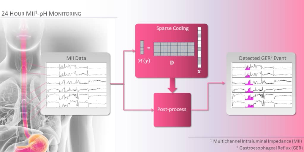
Multichannel Intraluminal Impedance data belonging to 26 individuals This dataset contains 174 episodes (2 minutes intervals) of Multichannel Intraluminal Impedance data belonging to 26 individuals. The dataset includes three variables, "IMPEDANCE", "BASE" and, "FLAG_GER" of the same size r*c. |
Thirteen 3D macular SD-OCT images obtained from eyes without pathologies using Topcon 3D OCT-1000 imaging system This dataset contains thirteen 3D macular SD-OCT images obtained from eyes without pathologies using Topcon 3D OCT-1000 imaging system in Ophthalmology Dept., Feiz Hospital, Isfahan, Iran. |
|
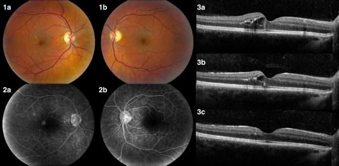
Dataset of Fully-labeled Diabetic Macular Edema OCT B-scans (associated with fluid and layer annotations) We enrolled twenty eyes from 19 patients with the diagnosis of diabetic macular edema (DME). All patients had clinical and OCT-based diagnosis of DME. OCT examinations were performed using Spectralis Spectral Domain-OCT for .the normal and DME patients |
Other Datasets
|
OCT data & Color Fundus Images of Left & Right Eyes of 50 healthy persons |
||
|
Data-set of corneal OCT taken from Heidelberg OCT imaging system (3D .mat data of 15 Subjects ) |
||


