
Thirteen 3D macular SD-OCT images obtained from eyes without pathologies using Topcon 3D OCT-1000 imaging system
This dataset contains thirteen 3D macular SD-OCT images obtained from eyes without pathologies using Topcon 3D OCT-1000 imaging system in Ophthalmology Dept., Feiz Hospital, Isfahan, Iran. The datasets are in mat format and are named “1” to “13”. The x, y, z size of the obtained volumes was 512 × 650 × 128 voxels, 7 × 3.125 × 3.125 mm3, voxel size 13.67 × 4.81 × 24.41 μm3(as shown in next Figure).
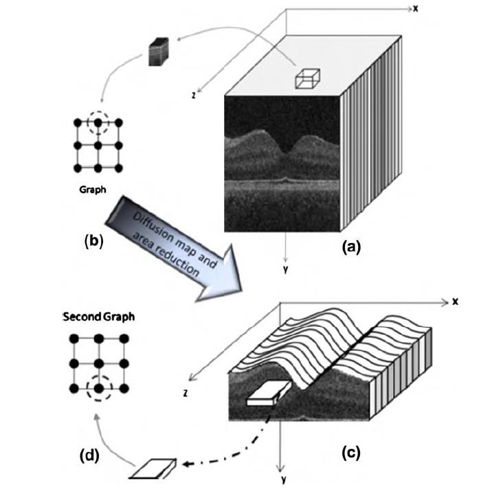

(a) Cubic windowing on 3D OCTs, (b) allocation of each node to a cubic portion of 3D OCTs, (c) narrowing the search area to pixels located between the 1st and 7th
surfaces and windowing of the new area, and (d) selecting thin horizontal cubic pixel boxes as the graph nodes.
The values of the proposed layer localization are provided for 12 boundaries shown in Figure 8 of the corresponding paper. The localization errors are reported on 10 randomly selected slices. The position of the mentioned slices are stored in another mat file named “randomImages”(the rows correspond to each dataset and the columns contain the traced slices).. Furthermore, the validation was based on manual tracing by two observers and mean value of the mentioned tracings are provided in mat files named “totalManual_1” to “totalManual_13”.
If you used this dataset, please cite the following papers: