|
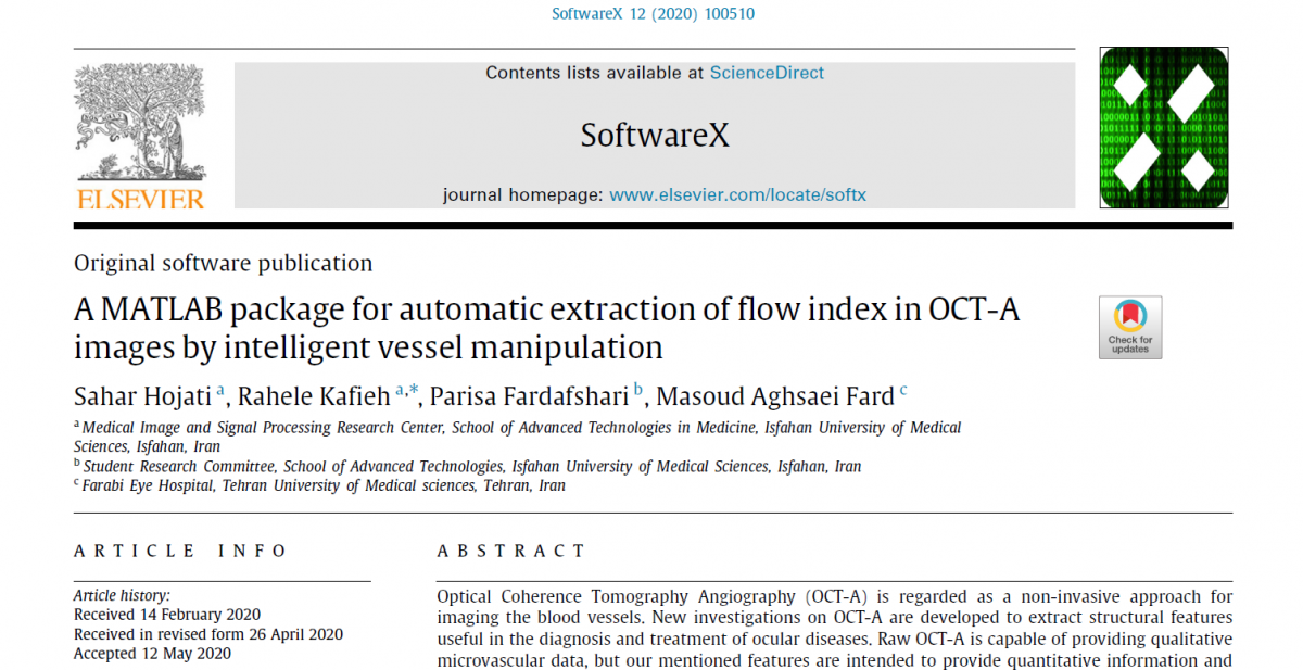
A MATLAB package for automatic extraction of flow index in OCT-A images by intelligent vessel manipulation |
|
|
Software Production 2020
|
|
|
Optical Coherence Tomography Angiography (OCT-A) is regarded as a non-invasive approach for imaging the blood vessels. New investigations on OCT-A are developed to extract structural features useful in the diagnosis and treatment of ocular diseases. Raw OCT-A is capable of providing qualitative microvascular data, but our mentioned features are intended to provide quantitative information and trustable comparison during follow ups and between different people. In this paper, we proposed an easy-to-use software to remove spurious blood vessel shadows and extract numerical features from OCT-A images. The features are expected to play an important role in the diagnosis and understanding of blood conditions. For this purpose, we used blood flow information as vessel density (VD) map (%) in a 4.5×4.5 mm rectangle scan centered on the optic disk for deep optic nerve data. Information from original image and image with small vessels were measured in whole-image (WI), peripapillary (PP) areas and theirs superior and inferior sectors. Furthermore, we also used blood flow information at parafoveal capillary as VD map in order to remove the projection artifact of superficial macular vessels from deep macular images, compatible with different analysis protocols for macula (3×3, 4×4, 5×5, 6×6, and 7×7 mm). Similarly, information from whole image and small vessels were measured in whole-image (Wi) and sector-based parafovea areas. |
|
|
Design & develop an OCT-based ocular health kiosk to diagnose Diabetic Retinopathy |
|
|
|
Avicenna Research Projects 2020
|
|
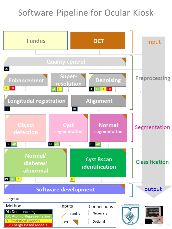
Diabetic retinopathy (DR) is revealed to be as one of the main complications of diabetes with prevalence of 95% and 60% in diabetic patients of type 1 and type 2 with over twenty years disease duration, respectively. This makes diabetes one of the leading phenomena for blindness and low vision.Vision loss due to DR is irreversible in most cases. However, risk of blindness can be reduced up to 95% by early detection and treatment. An annual eye exam is proposed for people with diabetes, because DR is not often associated with early symptoms. In addition, people with diagnosed DR may need more frequent eye exams for follow up. The regular DR screening routine leads to delayed diagnosis until progression to severe DR such as proliferative diabetic retinopathy (PDR) or diabetic macular edema (DME). People at risk of developing severe DR may need a comprehensive eye exam as frequently as every 2 to 4 months. Since current DR screening requires specialist equipment and expertise of the clinicians, only 35%-60% diabetic patients receive annual visit. By designing an accessible automatic ocular health kiosk, more frequent and scheduled screening of the diabetic patients will be provided, and early stages of preventable, treatable and potentially blinding disorders can be detected. |
|
|
3D-Printing of Resin-Based Parts with Magnetic Properties |
|
|
Choc Edge Ltd, United Kingdom 2020 |
|
|
cluding Fe3O4 nanoparticle inside printing ink can produce polymeric parts with para-magnetic properties. It is believed that applying magnetic fields to printing ink during printing can align the nanoparticles within the printed structure and improve its para-magnetic properties. In this project, we are studying surrounding printing nozzle with several different orientations of magnetic field and investigate the magnetic strength of final parts. |
|


|
lodGWAS: a software package for genome-wide association analysis of biomarkers with a limit of detection |
|
|
Prof. Harold Snieder Department of Epidemiology, University of Groningen, University Medical Center Groningen, Groningen, 2016 |
|
|
Genome-wide association study (GWAS) of a biomarker is complicated when the assay procedure of the biomarker is restricted by a Limit of Detection (LOD). Those observations falling outside the LOD cannot be simply discarded, but should be included into the analysis by applying an appropriate statistical method. However, the problem of LOD in GWAS analysis of such biomarkers is usually overlooked. ‘lodGWAS’ is a flexible, easy-to-use R package that provides a simple and elegant way for GWAS analysis of such biomarkers while simultaneously accommodating the problem of LOD by applying a parametric survival analysis method. |
|
|
Assessing the Underworld |
|
|
Engineering and Physical Sciences Research Council, United Kingdom The University of Sheffield 2016 |
|
|
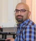
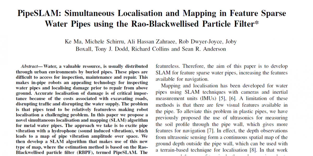
Water, a valuable resource, is usually distributed through urban environments by buried pipes. These pipes are difficult to access for inspection, maintenance and repair. This makes in-pipe robots an appealing technology for inspecting water pipes and localising damage prior to repair from above ground. Accurate localisation of damage is of critical impor-tance because of the costs associated with excavating roads, disrupting traffic and disrupting the water supply. The problem is that pipes tend to be relatively featureless making retal water pipes. The approach we take is to excite pipevibration with a hydrophone (sound induced vibration), which leads to a map of pipe vibration amplitude over space. We then develop a SLAM algorithm that makes use of this new type of map, where the estimation method is based on the Rao- Blackwellised particle filter (RBPF), termed PipeSLAM. The approach is also suited to SLAM in plastic water pipes using a similar type of map derived from ultrasonic sensing. We successfully demonstrate the feasibility of the approach using a obot localisation a challenging problem. In this paper we propose a novel simultaneous localisation and mapping (SLAM) algorithm for mcombination of experimental and simulation data. |
|
|
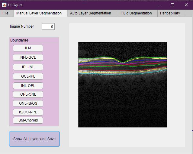
Livelayer: A Semi-Automatic Software for Segmentation of Layers and Objects in Optical Coherence Tomography Images |
|
|
Software Production 2020 |
|
|
The Ophthalmology Science has recently witnessed marked progress due to the advent of divergent imaging techniques, especially the Optical Coherence Tomography (OCT) which has caught many physicians’ attention for being exact, rapid, non-invasive and low-cost. Given these interesting features of OCTs as well as the capacity to display symptoms of a wide variety of eye diseases and neurological disorders, the need for OCT image segmentation and the corresponding data interpretation is felt more than ever before. In this paper, we wish to address this need and solve the difficulties associated with OCT image segmentation by offering a piece of handy, first-time software written in the MATLAB App Designer environment which helps researchers and clinicians to easily segment the images, save the numerical outcomes and send them for proper analysis. Serving an unambiguous user interface along with a unified platform in which all necessary functions have been incorporated graphically has made this software so unique that it could be recommended to anyone tending to work on ocular OCT layers and fluid segmentation. The software also accommodates a novel graph-based semi-automatic method, called “Livelayer” and designed for straightforward segmentation of retinal layers and fluids. |
|

|
Dynamic Registration and Intraoperative Navigation for Image Guided Spine Surgery |
|
|
Mirzakhani Research Projects 2020
|
|
|
Surgical navigation systems are used to assist surgeons in navigating within an intraoperative environment and in planning and guiding surgery. Similar to a car or mobile Global Positioning System (GPS), image guided surgery systems (IGS) use cameras or electromagnetic fields to capture and relay the patient’s anatomy and the surgeon’s precise movements in relation to the patient, to computer monitors in the operating room. Indeed, IGS is the use of a real-time correlation of the operative field to a preoperative imaging dataset that reflects the precise location of a selected surgical instrument to the surrounding anatomic structures. The accuracy of the IGS system is very crucial in localizing and positioning surgical instruments during surgery. Navigation surgery and Image-guided techniques have not been widely used in spinal surgery due to complicated patient registration process. It has been limited by several factors such as vertebral and intervertebral motions and inadequate immobilization of spine. In current techniques of spinal IGS, the surgical field is registered with a pre-operative computed tomography (CT), often obtained in a different spinal confirmation, or with intraoperative imaging such as fluoroscopy or intraoperative computed tomography (iCT). Intraoperative Stereovision (iSV) offers an alternative radiation-free method of registration to intraoperative CT (iCT). Intervertebral cumulative movements make the impossible use of skin-affixed fiducials and registration at the start of surgery could not compensate intervertebral motion between preoperative CT scans and intraoperative patient position. An automated registration procedure is proposed using iSV, which could accelerate adoption and improve outcomes, in addition to reducing costs, complexity, and x-ray dose associated with current spine surgical guidance methods. A total of four phases (phantom, cadaver, live animal, and human subjects) will be performed. The primary purpose of this study is to evaluate the performance of the iSV methods we previously developed in the setting of image-guided cranial surgery for rapid intraoperative patient registration and compensate intervertebral motion in open spinal surgery procedures with minimal operator intervention. If this proposal would be successful, these findings support the clinical feasibility of iSV to offer radiation-free, noninvasive imaging technique as the basis for establishing accurate, efficient and robust image-guidance and navigation during spinal surgery, and therefore, its potential to further increase the adoption of image-guidance in this surgical specialty. |
|

|
A feasibility study to develop an OCT-based ocular health kiosk to diagnose Diabetic Retinopathy |
|
|
Iran-Switzerland Research Seed Money Grant Basel University 2019 |
|
|
Diabetic retinopathy (DR) is revealed to be as one of the main complications of diabetes with prevalence of 95% and 60% in diabetic patients of type 1 and type 2 with over twenty years disease duration, respectively. This makes diabetes one of the leading phenomena for blindness and low vision. Vision loss due to DR is irreversible in most cases. However, risk of blindness can be reduced up to 95% by early detection and treatment. An annual eye exam is proposed for people with diabetes, because DR is not often associated with early symptoms. In addition, people with diagnosed DR may need more frequent eye exams for follow up. The regular DR screening routine leads to delayed diagnosis until progression to severe DR such as proliferative diabetic retinopathy (PDR) or diabetic macular edema (DME). People at risk of developing severe DR may need a comprehensive eye exam as frequently as every 2 to 4 months. Since current DR screening requires specialist equipment and expertise of the clinicians, only 35%-60% diabetic patients receive annual visit. By designing an accessible automatic ocular health kiosk, more frequent and scheduled screening of the diabetic patients will be provided, and early stages of preventable, treatable and potentially blinding disorders can be detected. So, the specific aims regarding to designed ocular health kiosk would be: Aim1. Feasibility study of developing the hardware to have an auto-focus auto-capture OCT image acquisition system without operator intervention Aim2. Designing and tuning the automatic OCT image classification software for DR detection Aim3. Evaluating the accuracy and false positive ratio (FPR) of ocular health kiosk in comparison to clinical judgment on Screening and Follow-up of DR. |
|
|
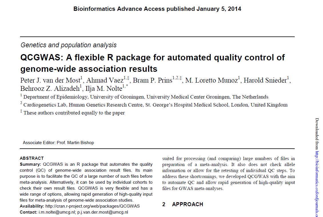
QCGWAS: A flexible R package for automated quality control of genome-wide association results |
|
|
Prof. Peter J. van der Most Department of Epidemiology, University of Groningen, University Medical Center Groningen, Groningen, 2014 |
|
|
QCGWAS is an R package that automates the quality control of genome-wide association result files. Its main purpose is to facilitate the quality control of a large number of such files before meta-analysis. Alternatively, it can be used by individual cohorts to check their own result files. QCGWAS is flexible and has a wide range of options, allowing rapid generation of high-quality input files for meta-analysis of genome-wide association studies. |
|


| Automatic Multifaceted Matlab Package for Analysis of Ocular Images (AMPAO) | |
|
Software Production 2019 |
|
| Ocular imaging can be found amongst the most prevalent and popular techniques that provide information about diseases affecting the visual system. Such information may be incorporated in applications beginning from diagnosis, through treatment, and extending to follow-ups. Optical Coherence Tomography (OCT) and enface ocular imaging like fundus and scanning laser ophthalmoscopy (SLO) are the most approved modalities since they are non-invasive, highly informative and comparatively inexpensive. There are already some established software for ocular image analysis; however, to be of use in real applications, the data analysis should be gathered in a platform that enables the data to flow through the software components. The principal goal of the software introduced in this paper is to accept different data formats, and to pipeline the data to different ocular analysis techniques. Furthermore, AMPAO permits use of each algorithm individually. Additionally, the software is flexible and could employ other analysis algorithms to be embedded in different sections. | |
|
Developing Micro-Scale Network via Near-Field Electrospinning |
|
|
Choc Edge Ltd, United Kingdom and China Wuhan University 2019 |
|
|
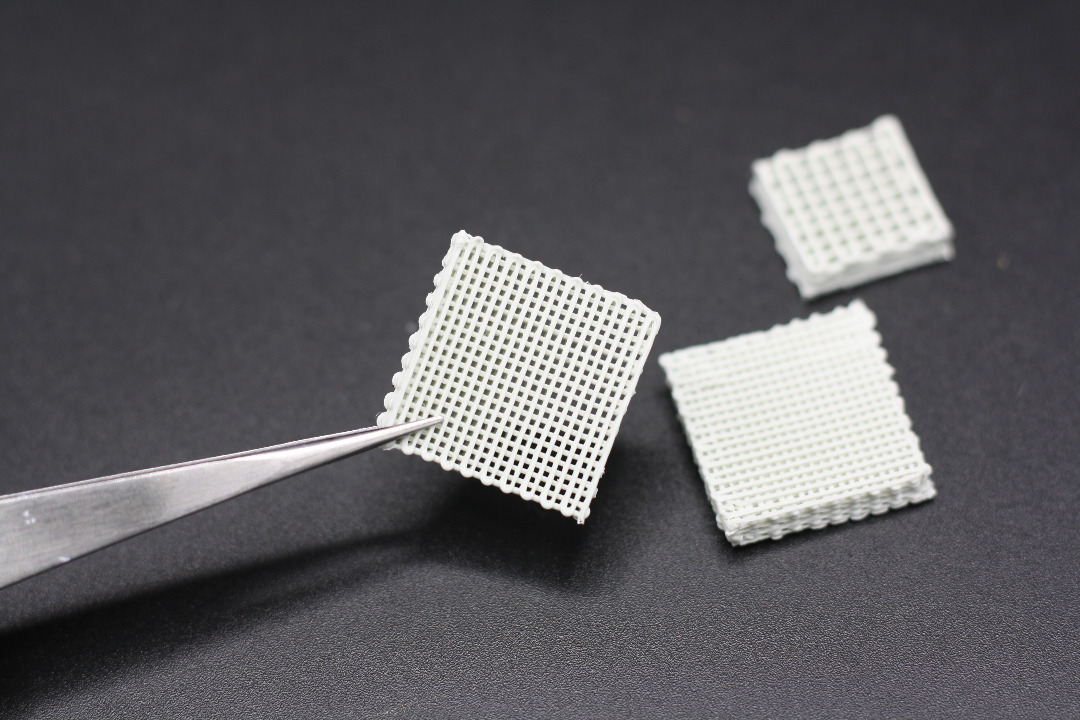
One of the most advanced manufacturing technique in the field of electrospinning is called “Near-Field Electrospinning” which can produce extremely precise pattern of deposited micro- or nano-scale fibers according to a desirable predefined pattern. In this project we are studying the physics behind fibre formation during electrospinning process and also try to produce Collagen-PCL scaffolds and via near-field electrospinning compare their properties with traditionally produced Collagen-PCL scaffolds. |
|
|
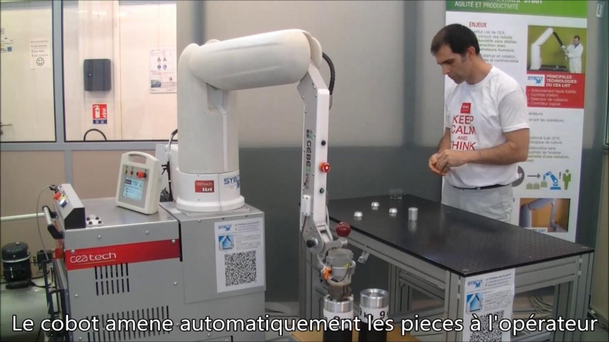
ADN4SE: Embedded systems kernel & development for collaborative robotics |
|
|
Agence Nationale de la Recherche, France Laboratoire de Robotique Interactive, CEA-List, Saclay, France 2015 |
|
|
The ADN4SE project aims at understanding how humans perform the manipulation of objects in order to replicate grasping and skilled in-hand movements with an anthropomorphic artificial hand, and thereby move robot grippers from current best practice towards more autonomous, natural and effective articulated hands. The goal is to endow the proposed robotic hand with advanced perception capabilities, high level feedback control and elements of intelligence that allow recognition of objects and context, reasoning about actions and a high degree of recovery from failure during the execution of dexterous tasks. |
|

|
Design and application of Bandlet on Graph for image denoising |
|
|
Prof. Gozde Unal TUBITAK Istanbul Technical University, Turkey 2014 |
|
|
We introduce a new image modeling method by getting benefit from both sparsity and multiscale characteristics of transform-domain modeling, along with the geometrical representation of the graph-based models. The proposed method is named bandlet on an oriented graph (BOG) and improves directional selectivity property of the bandlets. The conventional wavelet in the bandlet design is substituted with a new non-orthogonal wavelet. The replaced wavelet is defined on a graph. In order to adjust the orientation of the wavelet atoms with the corresponding edges in the image pixels, a directed graph is constructed. The resultant wavelets in discrete scales can be considered as a frame and are created to build a tight frame. To show the effectiveness of this new atomic representation, we demonstrated the performance of the new model in noise alleviation of the optical coherence tomography (OCT) images (from the retina) and microscopic images. Denoising results on OCT are reported on 72 slices, selected arbitrarily out of OCT dataset from Topcon device. The combined method provided an enhancement of contrast to noise ratio (CNR) (from 27.82 to 30.11), and improvement of the equivalent number of looks (ENL) (from 2183.26 to 2217.37) over the state-of-the-art in OCT noise reduction. In the denoising of microscopic images, PSNR improvement (from 26.33 to 35.24) over the original image is shown along with the improvement in next steps of feature extraction. |
|
|
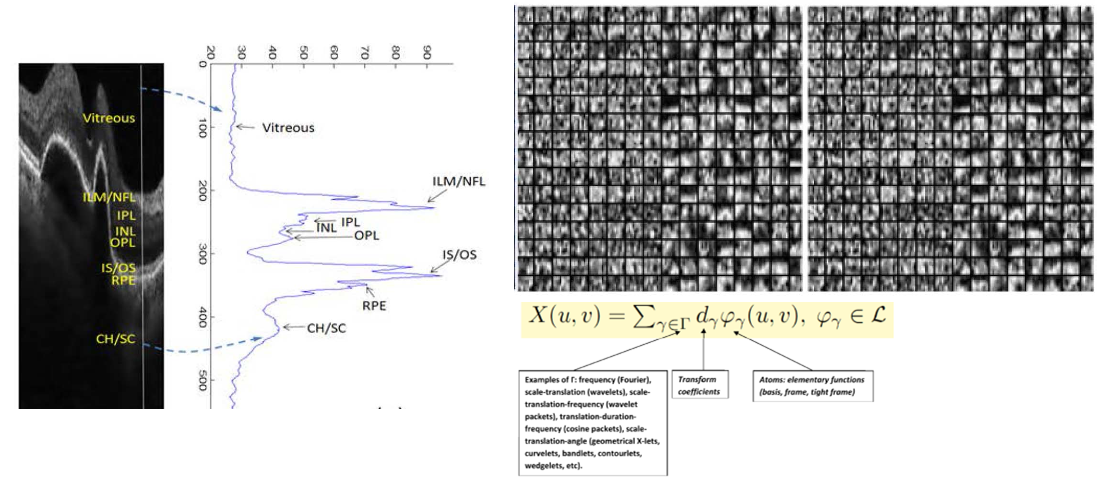
OCT-let: Designing an Optimum Sparse Representation for Ophthalmic Optical Coherence Tomography Image Analysis |
|
|
University of Gottingen Germany 2019 |
|
|
The research program proposed in this application aims to analyze Optical Coherence Tomography (OCT) images using sparse representations in suitable dictionaries. OCT is a new imaging technique to provide information about cross-sectional structures of an object. One of the most significant applications of OCT in clinical application is in the field of ophthalmology and has become an important tool to obtain detailed images from within the retina. The foundation of any signal processing technique is modeling which can be facilitated in transform domains using suitable dictionaries. The transform domain models are broken into data-adaptive and non-data adaptive transforms. These data representations are then applied in stochastic, deterministic, geometric or energy based models and usually lead to a regularization functional, where sparsity of the data in the fixed dictionary is taken as a prior information. The proposed research aims to take advantage of various transform-based modeling approaches, allowing better analysis of ocular OCTs. It is not clear a priori, if we can take one of the established non-adaptive systems or need to construct new dictionaries that respect the properties of OCT images implemented by the underlying physics. We also need to decide whether to take a data-adaptive or non-adaptive dictionary to produce optimal atomic representations can be used for ocular OCT images. In a second step, we want to combine the discovered dictionary to improve current approaches for OCT classification, OCT segmentation and OCT restoration. So, the outcomes of this research would be:
|
|
|
SPY: Surveillance imProved sYstem |
|
|
ITEA2, European Union Institut d’Electronique Fondamentale, Universite Paris-Sud, France 2014 |
|
|
The SPY project is creating an intelligent surveillance and situation awareness framework adapted to a mobile environment. The new framework will improve the use of wireless infrastructures and mobile sensors to help professional end users in the field, including the police and fire brigades in their surveillance and rescue tasks. |
|

|
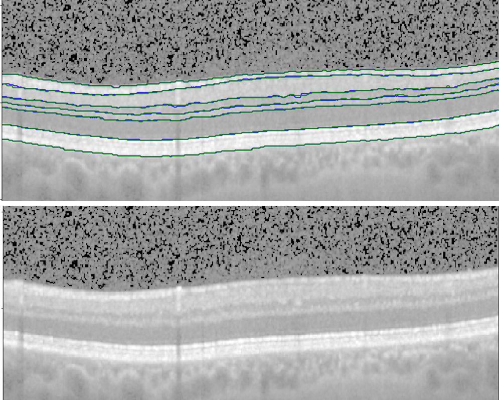
Berlin-Deep Algorithm for retinal segmentation in neurodegenerative diseases |
|
|
Prof. Alexander Brandt EINSTEIN Forum Neurocure Clinical Research Center (NCRC), Charite Medical University, Berlin, Germany 2018 |
|
|
Our method is a cascaded two-stage CNN model which incorporates the location awareness by adding the prior knowledge about inter-retinal layers. Our method includes following steps:, (a) first CNN for extraction of prior location knowledge, (b) second CNN based on inclusion of location knowledge from previous step in class-attended multi-dimensional U-net, (c) training on first dataset and fine tuning on second dataset, (d) extraction of inter-retinal boundaries from retinal layer information after applying simple morphological image processing techniques to reject anatomical infeasible results. We have already designed an accurate and clinically applicable deep learning method for OCT segmentation. The method was trained on OCTs from 2700 eyes (130593 Bscans), cross-sectionally gathered from healthy controls and people suffering from MS and NMO. The gold standard delineation on this dataset was achieved using available segmentation tools and was manually corrected in some cases. We then fine-tuned the network using OCTs from 48 eyes (2322 Bscans) from healthy controls and people suffering from MS and NMO. The second set had very precise gold delineation which was double checked for any possibly wrong segmentation. The method was then tested on 30 OCTs (1451 Bscans) with very accurate gold segmentation (similar to second dataset). Furthermore, we evaluated the method on 38 OCTs (1837 Bscans) from NMO patients with extremely thin layers which makes the segmentation more difficult. |
|
|
ID2U: Dexterous single-use instrument for laparoscopic surgery |
|
|
Agence Nationale de la Recherche, France Institut des Systemes Intelligents et Robotique, Sorbonne University 2010 |
|
|
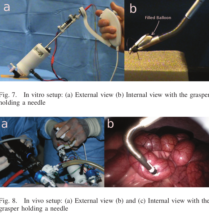
Minimally invasive surgery (MIS), which typically involves endoscopic camera and laparoscopic instruments may seem to be the ideal surgical procedure for its apparent benefits. However, in comparison to open surgeries, the spatial and mechanical tool limitations posed on surgeons are so high that often MIS is foregone for complex cases and even when it is possible, the procedure requires a high dexterity, caliber, and experience from the surgeon. Particularly, suturing procedure through MIS is known to be extremely challenging. We are working toward the development of a robotic hand-held surgical device for laparoscopic interventions that enhances the surgeons’ dexterity. The instrument produces two independent DOFs, which is sufficient for enabling MIS suturing procedure in vivo. The end-effector’s orientation is controlled by an intuitive and ergonomic controller and its position is controlled directly by the surgeon. Different control modes, handles, and end-effector kinematics are primarily evaluated using a virtual reality simulator before choosing the best combination. A proof-of-concept prototype of the device has been developed. |
|