
Introduction
Today, applications of Artificial Intelligence and Machine Learning have affected human lives in a wide range of issues and increased the efficiency of activities. Using AI methods in the field of medicine and for processing of medical images has received much attention and has improved the efficiency of image processing. Therefore, in order to emphasize the importance of familiarizing with the applications of AI in the field of radiology we have decided to hold a competition for quantitative processing of radiological images in the 29th National Conference and the 7th International Conference of Biomedical Engineering of Iran.
problem statement
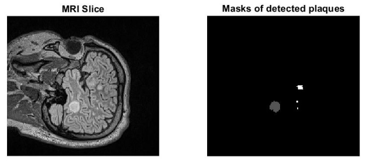
Multiple Sclerosis (MS) is an autoimmune disease involving the central nervous system in which the body's immune system destroys the myelin sheath of neurons in the brain and spinal cord. These areas appear as plaques in MRI. These plaques are usually not visible in T1 phase images in MRI and are seen as white areas in T2 phase and FLAIRE in MRI. In MRI protocol with the injection of radioactive substances (gadolinium), these plaques are seen as bright incomplete rings in T1. These plaques can be in the Periventricular, White Matter, Juxtacortical and Infratentorial areas of the brain. Determining the number and location of plaques is very important in the certainty of MS diagnosis according to the McDonald index. An example of an MRI image showing the location of the plaques has been illustrated in the following figure.
Determination of the number and location of plaques has been done visually so far, and this has brought limitations in determining the exact number and location of plaques. Accordingly, the use of image processing methods and AI in this field can lead to an increase in the accuracy and quality of image evaluation and consequently helps to more accurate diagnose of this disease.
Based on the above explanation, in this competition we aim to determine the best and most qualitative algorithms for detecting and locating plaques in the brain.
Procedure
In order to run this competition, 75 MRI images of MS patients have been prepared. Imaging was done with an MRI machine (Siemens company, Germany, 1.5 Tesla MRI Siemens Avanto scanner system Siemens, Henkestr Erlangen). The patient is placed supine on the bed and the patient's head is placed in a twelve-channel coil for head imaging and the image is prepared. The obtained images have NIFTI format and the size 236x256x160.
In the first stage, 55 images will be provided to the participants. Participants need to design a new algorithm to detect brain plaques in juxtacortical, white matter, periventricular and infratentorial areas. Each group must report the results based on the explanations that will be presented below. The received algorithms and results will be reviewed the best presented algorithms will go to the second stage.
In the second stage, the remaining 20 test data will be provided to the selected groups and the best results in this stage will be the winners.
How to participate in the contest
Participation is open to all. Each participating team will consist of a maximum of 3 people. Contest organizers are not allowed to participate.
Interested groups must complete the registration form and send it to misp@mui.ac.ir. If the information is confirmed, the data link will be sent to them.
Download the registration form
How to present the results
It is necessary for each participating group to send the results obtained from the first stage via email to the following address.
misp@mui.ac.ir
Submitted results should be in the form of a zip file containing the obtained mask for the plaques in each image and an article containing a full description of the implementation method, the proposed algorithm, and the obtained results.
important dates
Deadline for registration and receiving data:4 November 2022
Deadline for sending preliminary results: Extension up to 29 November 2022
Announcement of preliminary results and selected people of the first stage: 11 December 2022
Holding the second stage and announcing the names of the selected people: 21 December 2022
Prizes for selected people:
The best works will be invited to be published in the Journal of Medical Signals and Sensors indexed in Pubmed, Scopus and Web of Science and an article comparing the best results will be published.
The organizers will evaluate the quality of the results for publication.
|
Name |
Degree |
|
Adress |
|
Iman Adibi |
Specialized Doctorate in Medicine / Neurological Diseases |
Faculty of Medicine - Isfahan University of Medical Sciences |
|
|
Hossein Rabbani |
Phd of Biomedical Engineering |
h_rabbani@med.mui.ac.ir |
Medical Image and Signal Processing Research Center - Isfahan University of Medical Sciences |
|
Farnaz Sedighin |
Phd of Electrical Engineering |
f.sedighin@amt.mui.ac.ir |
Medical Image and Signal Processing Research Center - Isfahan University of Medical Sciences |
|
Maryam Ansarian |
Master of Biomedical Engineering |
maryamansarian70@yahoo.com |
Medical Image and Signal Processing Research Center - Isfahan University of Medical Sciences |
|
Ghazal Zandieh |
Doctor of Medicine |
|
|
|
Neda Ramezani |
Doctor of Medicine |
|
|
|
Safieh Danesh |
Master of Anatomy |
Samiradm1362@gmail.com |
|
|
Vahid Shayghan nejad |
Specialized Doctorate in Medicine / Neurological Diseases |
v.shaygannejad@gmail.com |
Faculty of Medicine - Isfahan University of Medical Sciences |
|
Fariba Ashtari |
Specialized Doctorate in Medicine / Neurological Diseases |
fashtari231@gmail.com |
Faculty of Medicine - Isfahan University of Medical Sciences |
For more information, please send email to misp@mui.ac.ir or use phone number 031-37925252.